Digital anatomy table gives RIT’s physician assistant BS/MS program a high-tech teaching tool
Traci Westcott/RIT
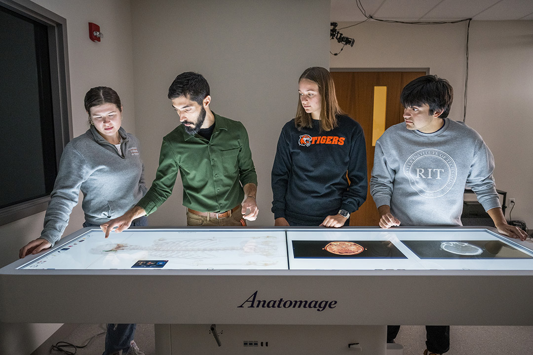
A digital anatomy table gives the RIT physician assistant BS/MS program a new high-tech teaching tool. Physician assistant lecturer Christopher Montanaro, second from left, works with his students Katrina Breen, left, Kaitlyn Bockelman, third from left, and Justin Koplinka.
A digital anatomy table programmed with dynamic medical simulations is changing how RIT physician assistant students learn about the human body.
Three-dimensional simulations modeled on real cadavers allow students to understand how the body functions as a system and deteriorates from disease.
The simulated cadaver table manufactured by Anatomage Inc. arrived early in the fall semester, and students are already using it to supplement their courses.
Traci Westcott/RIT
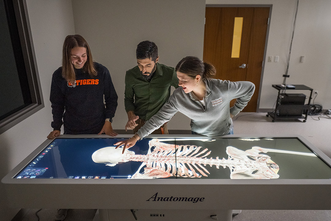
RIT physician assistant lecturer Christopher Montanaro, center, quizzes physician assistant students Kaitlyn Bockelman, left, and Katrina Breen, right, on a digital anatomy table.
Justin Koplinka, a third-year PA student from Fairport, N.Y., is learning about the heart in his clinical medical sciences class and is excited to use the Anatomage table to help him understand how the muscle functions and how to read EKG signals that trace the electrical pathways through the heart.
Each week, Koplinka and his classmates study hundreds of static medical slides from two-dimensional photographs and drawings. The dynamic medical visualization will help them understand the context, he said.
“It’s helpful to be able to visualize everything and to connect what we’re learning in pathology class to what we’re learning in clinical medical class,” Koplinka said. “I could use the table to see exactly how the heart is working and what the EKG would look like, so it’s a way to visualize all the different heart abnormalities.”
The equipment arrived early in the fall semester and physician assistant lecturer Christopher Montanaro ’10 (physician assistant) has immersed himself in learning how to use it himself and then teach faculty and students. He has programmed the table with scenarios from the software’s library of actual medical cases that support the PA program’s curriculum.
Students can download the mobile app and use the software on their cell phones and tablets. Montanaro encourages them to explore the learning modules, tests, and games, which can provide refreshers, especially for the fifth-year students doing their clinical internships.
“When they’re about to go on a clinical rotation, say surgical rotation, and they know they will be removing a gallbladder on a patient, for instance, they can go to the GI section and review it in detail and see it in 3D,” Montanaro said. “If they’re about to go on a big case, they can contact me and I can show them on the Anatomage table.”
The table will be in full use during the spring semester. Physician assistant students enrolled in Advanced Gross Anatomy will alternate between the cadaver lab to observe dissected bodies and the Anatomage table to practice clinical scenarios.
RIT’s cadaver lab can accommodate six specimens obtained from the University of Rochester Medical Center. The lab supports the human gross anatomy course taught by James Perkins, head of RIT’s Department of Medical Sciences, Health, and Management in the College of Health Sciences and Technology. Medical illustration majors take the course in the fall, and biomedical sciences and physician assistant students take it in the spring.
“The standard for learning human anatomy is the cadaver lab,” Montanaro said. “The Anatomage table doesn’t replace the cadaver lab, but supplements it.”
The Anatomage table is a large touch screen computer set into a waist-high table that can adjust into an upright position for a class. Images on the screen can be manipulated to change orientation and cross-sectional view.
The Anatomage software package comes with four adult cadavers modeled on actual people who have donated their bodies to science. Each body has been reconstructed to simulate a healthy form and to illustrate the transition to a diseased state down to the blood vessels.
Regional dissections based on many other cadavers show how different anatomy functions. Students learning about the cardiac system, for instance, can compare healthy and diseased hearts and corresponding EKG signals. Simulations like this help students synthesize and apply their understanding.
“When we’re teaching medicine, the mantra is ‘structure leads to function,’ and when the structure breaks down, the function breaks down,” Montanaro said. “The Anatomage table can help students start to think through it on their own. Any tool we can get to help us develop that deeper understanding, not just by memorizing, is critical.”
In addition to the cadavers and regional anatomy, the Anatomage table includes a pregnant woman to simulate child labor and birth.
“We were looking at the table right before our women’s health exam,” said Katrina Breen, a fourth-year physician assistant student from Dover, N.H. “The live birth shows all the stations throughout the birthing process. I think seeing it in a 3D format and having it be dynamic was helpful for me.”
The anatomy table also gives students context when learning to read two-dimensional medical imaging. They can pull up scans on the screen next to the body or regional dissection.
“We’re in diagnostic imaging, and it’s really interesting to see the different cuts and views,” said Kaitlyn Brockelman, a fourth-year physician assistant student from Cleveland, Ohio. “These are from real cadavers, so being able to see the different views of the pathology is very interesting. You can see the different layers of the anatomy.”
Koplinka thinks the Anatomage table will make a difference for students in the rigorous physician assistant program. Students must learn as much as they can about medicine before their fifth-year clinical rotations begin.
“A lot of us are visual learners, so having something like this at our disposal, even for a quick review, will help us a lot,” Koplinka said. “I am excited to use the anatomy table more.”


