Anatomy, Histology, and Pathology
Illustrations of anatomy, histology (cellular structure), and pathology. All created in Adobe Photoshop.
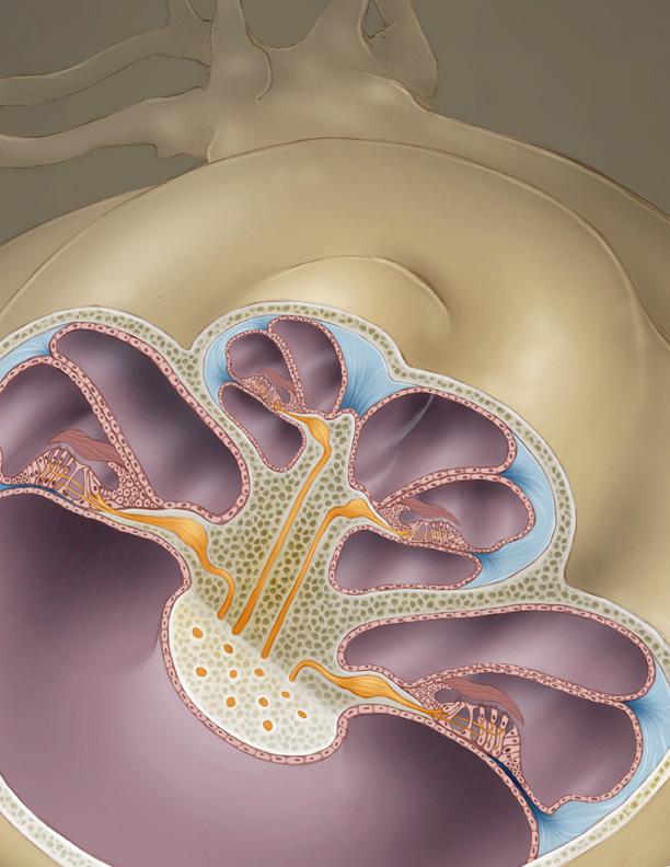
Organ of Corti. Aly Webster
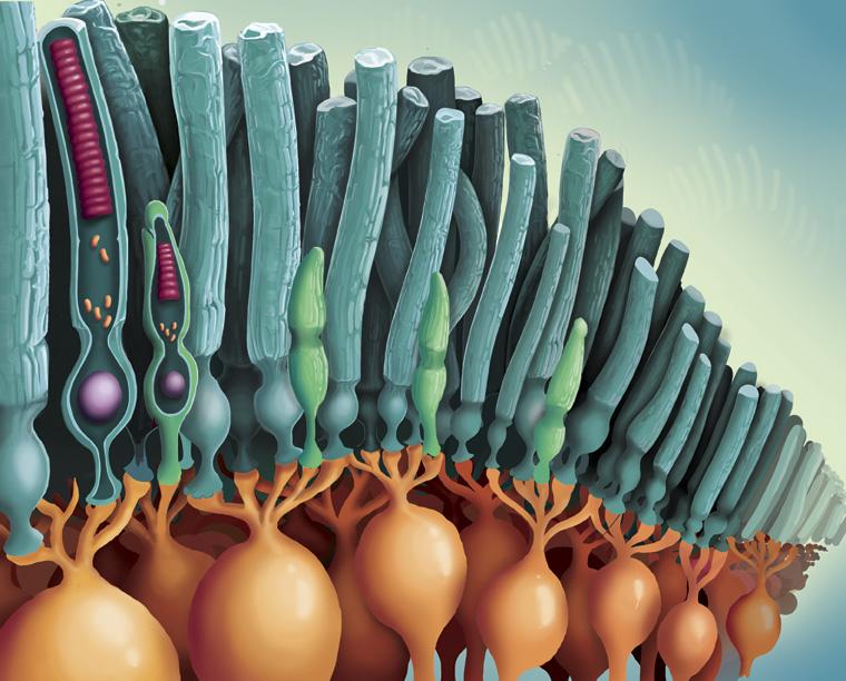
Cellular structure of the retina. Will Lawson
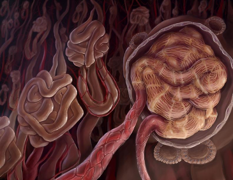
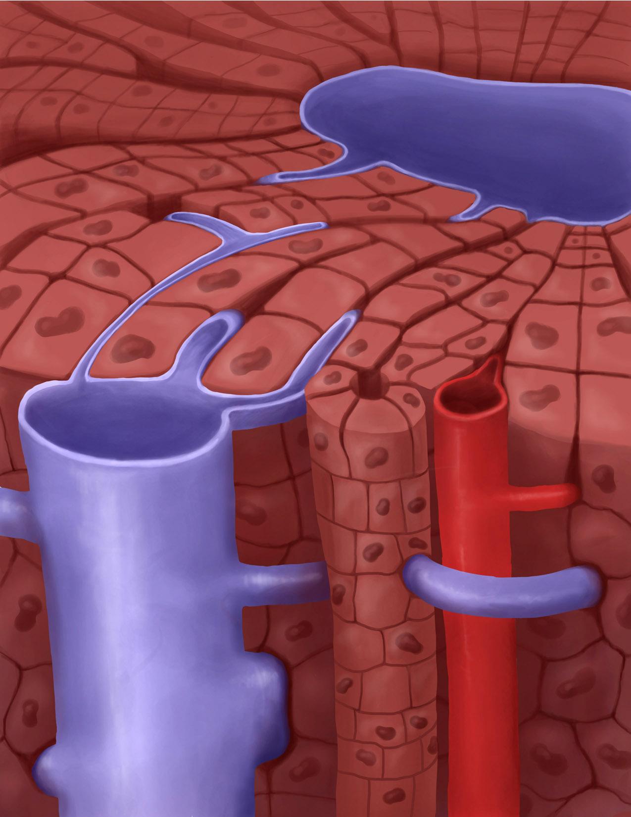
The glomerulus. Hannah Ely
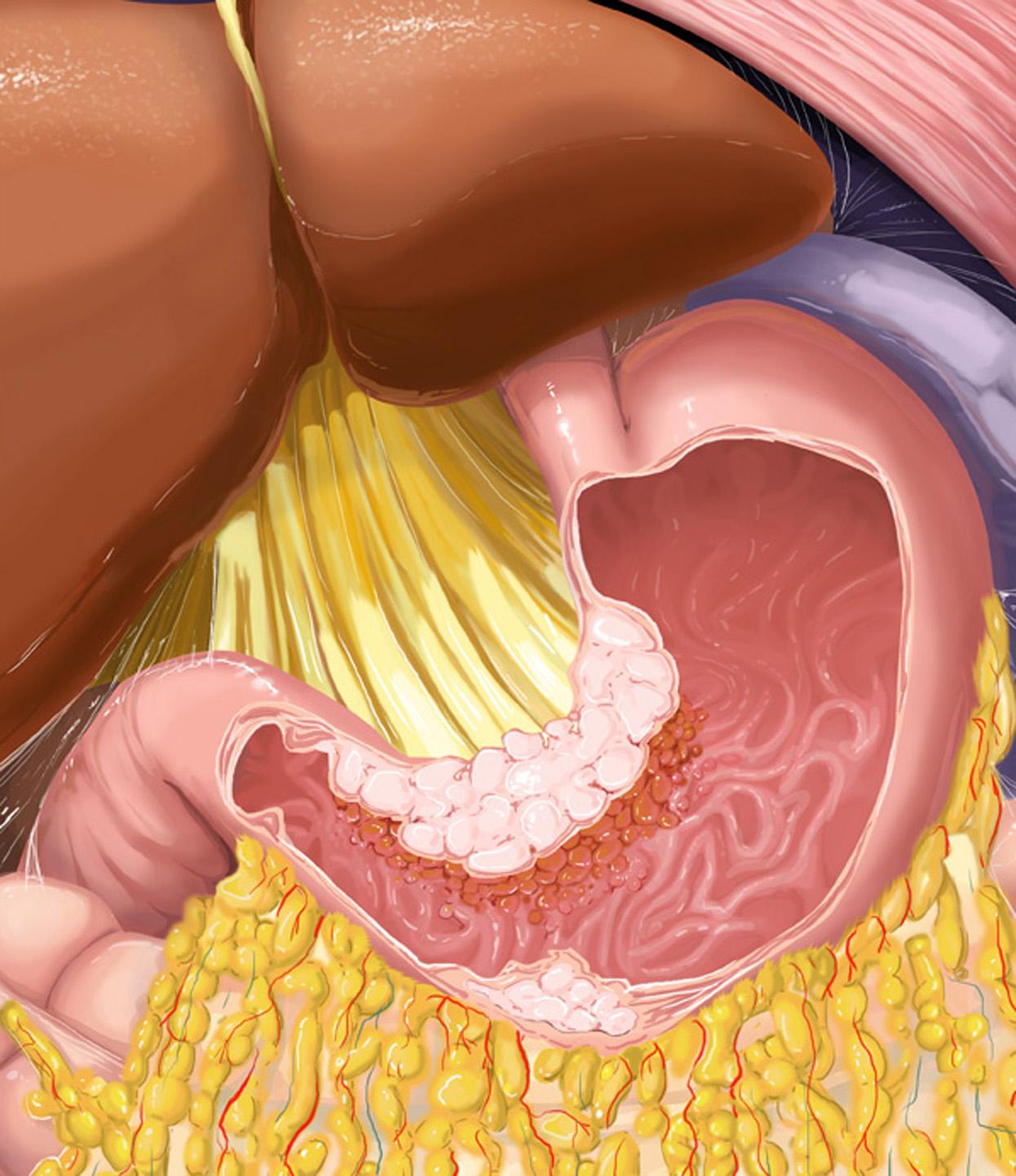
Adenocarcinoma of the stomach. Charlotte Cullen
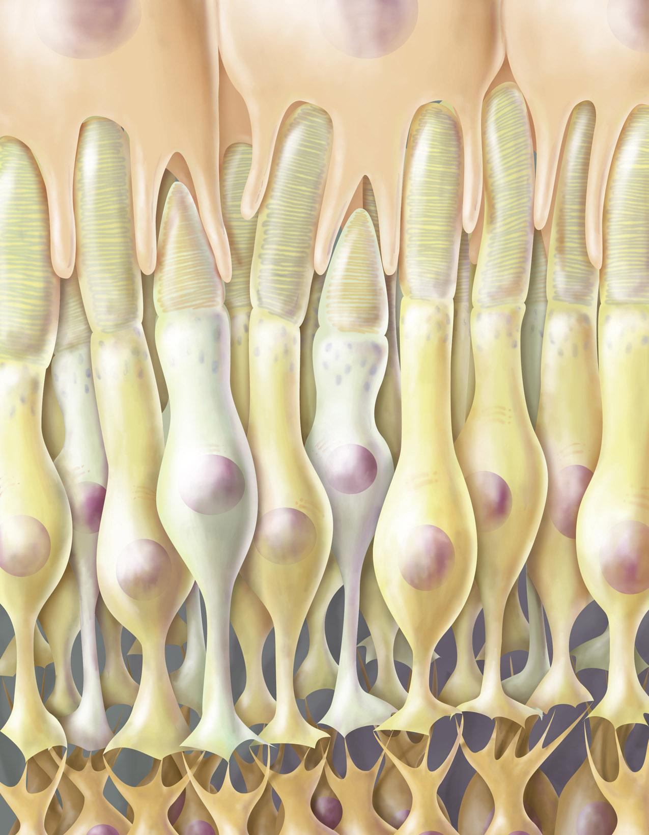
Cellular structure of the retina. Libby Lamb
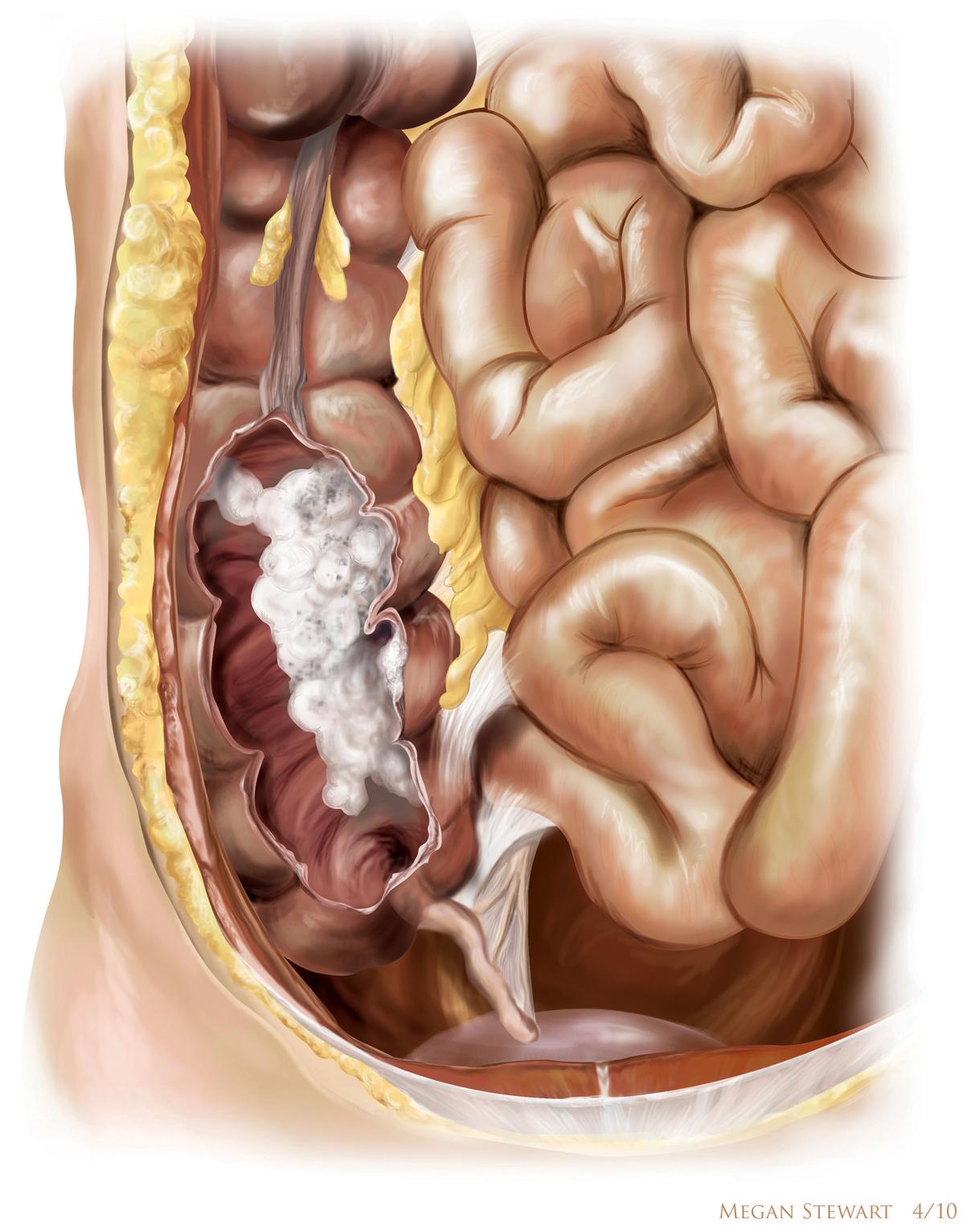
Adenocarcinoma of the colon. Megan Stewart
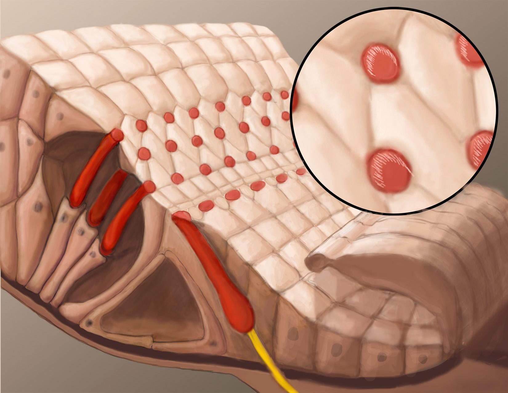
Cellular structure of the Organ of Corti in the inner ear. Siobhan McQueen
Cellular structure of the liver. Jackie Nee.
More Featured Profiles and Work
Donald Arday
Faculty

Michael Amy
Faculty

Robert Dorsey
Faculty
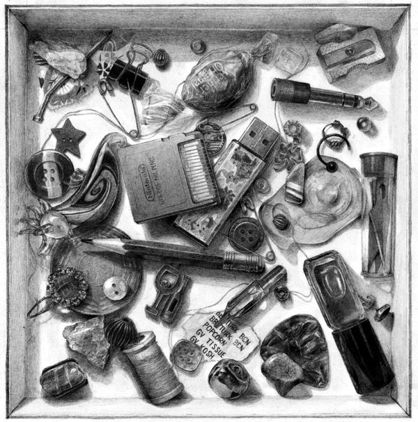
Foundations Drawing
Student