Engineering researchers receive NSF funding to develop computational tools to monitor ablation therapy on cardiac tissue
Data-driven platform being developed is needed to assess, guide and offer better visualizations of tissue response to thermal therapies
RIT
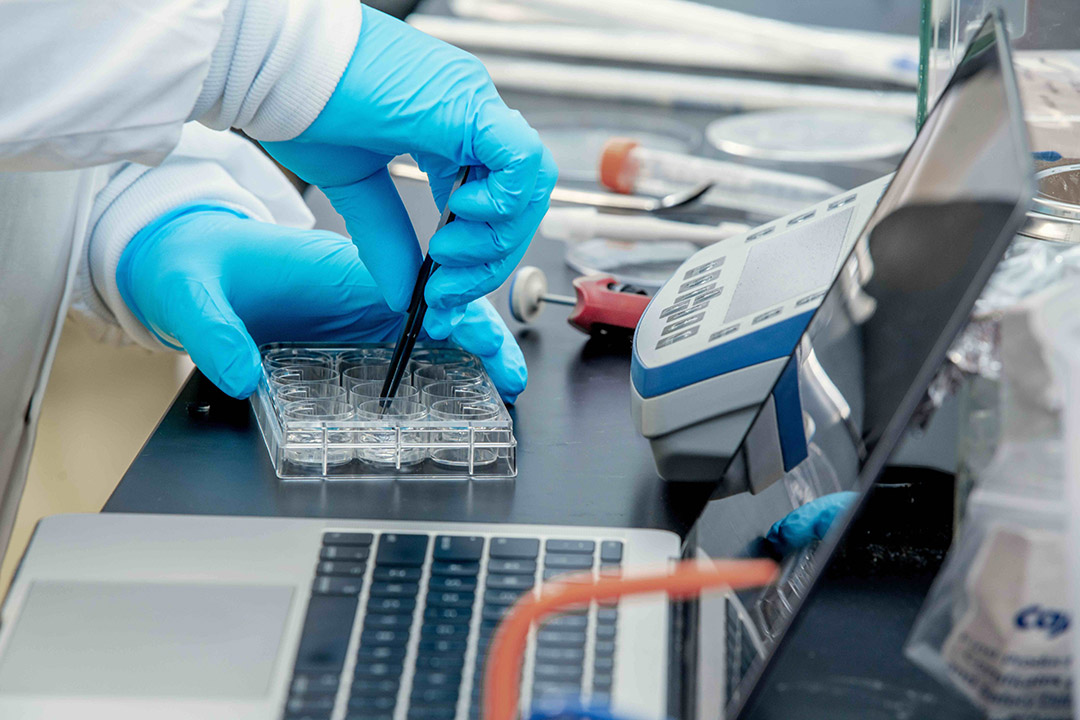
RIT engineering researchers are studying the effects of thermal ablation therapy on cardiac tissue to improve arrhythmia treatments.
Researchers at RIT are developing non-invasive technology that will better assess cardiac tissue response to thermal energy, a common therapy approach for both cancer and cardiac arrhythmia treatments.
Cristian Linte, a professor of biomedical engineering, is leading a cross-disciplinary team that was recently awarded National Science Foundation Collaborative Research funding to further develop an interactive, computing platform to characterize and monitor thermal cardiac tissue ablation therapy.
Ablation is a medical technique using heat, extreme cold, or chemical interventions to destroy abnormally functioning, diseased tissues. While effective, understanding how ablation affects healthy surrounding tissue has had limited study.
RIT
RIT biomedical engineering professor Cristian Linte
“Understanding thermal effects on biological tissues is crucial for many biological applications, including thermal therapies aimed at destroying pathological cells while preserving surrounding healthy tissue. However, there are no feasible means to monitor tissue response to thermal energy to characterize and visualize ablation patterns to ensure reversible injury and avoid incomplete ablation,” said Linte, an expert in medical imaging and image-guided technologies in RIT’s Kate Gleason College of Engineering.
Modern, minimally invasive ablative treatments for some cancers or heart rhythm disorders rely on the use of heat delivered to tissue in the form of radiofrequency energy to destroy a cancerous tumor or diseased tissue in the heart that causes rhythm abnormalities. The process blocks irregular heart signals and is a means to restore standard heartbeat patterns. Ablation provides an effective and less invasive option, but as many as 50 percent of ablation patients experience disease recurrence due to incomplete tissue ablation, Linte explained.
“Successful treatment requires continuous ablation patterns that induce permanent thermal damage to the tissue. However, temperature measurements inside the heart are invasive and unfeasible,” he said. “Unfortunately, routine medical imaging provides no quantitative appraisal of the induced thermal damage. Traditional computational tools used to study ablation are prohibitive for intra-operative use. There are no effective means to quantify heat transfer in biological tissue to interactively characterize and visualize thermal ablation lesions.”
RIT
RIT mechanical engineering professor Satish Kandlikar.
As part of the research team, Linte will be joined by Satish Kandlikar, RIT professor of mechanical engineering, and Suzanne Shontz, professor of electrical engineering and computer science at the University of Kansas, experts in heat transfer and computational bioengineering respectively.
Combining a cross-disciplinary approach, the team will explore heat transfer theory, image computing and visualization, biomedical modeling and simulation to build, model and quantify tissue response to thermal energy.
“Experimental validation is essential to confirm analytical and numerical models, especially in biological systems,” said Kandlikar who leads RIT’s Thermal Analysis and Microfluidics Lab. He will focus on the propagation of thermal energy and provide a temperature map of tissue during the ablation process in simulated tissue material that mimics biological tissue at the cardiac wall. The experimental data will be used to further refine the models developed during this study.
“Since knowledge of tissue temperature distribution is critical to accurately predict thermal effects, computationally efficient models that integrate engineering heat transport mechanisms alongside tissue pathophysiology are needed to quantify thermal energy effects on tissues and interactively characterize, visualize, and monitor induced thermal injury,” said Shontz, who also serves as the University of Kansas associate dean of research.
Shontz and Linte have collaborated on an earlier diagnostic project, also funded by the NSF, and similar in its development of computing and engineering models to appraise biomechanical heart functions.
This current project is already contributing new knowledge in scientific computing, engineering, and physiological modeling by helping to characterize thermal injury delivered to heart tissue during therapy.
“This research has the potential to reshape biological, engineering and material science applications that involve heat transfer modeling and heat flow characterization by enabling, for the first time, intra-operative, quantitative monitoring of heat transfer and thermal effects,” said Linte.


