Scientific Illustration
These illustrations were created in the Medical Illustration program’s Scientific Visualization course.
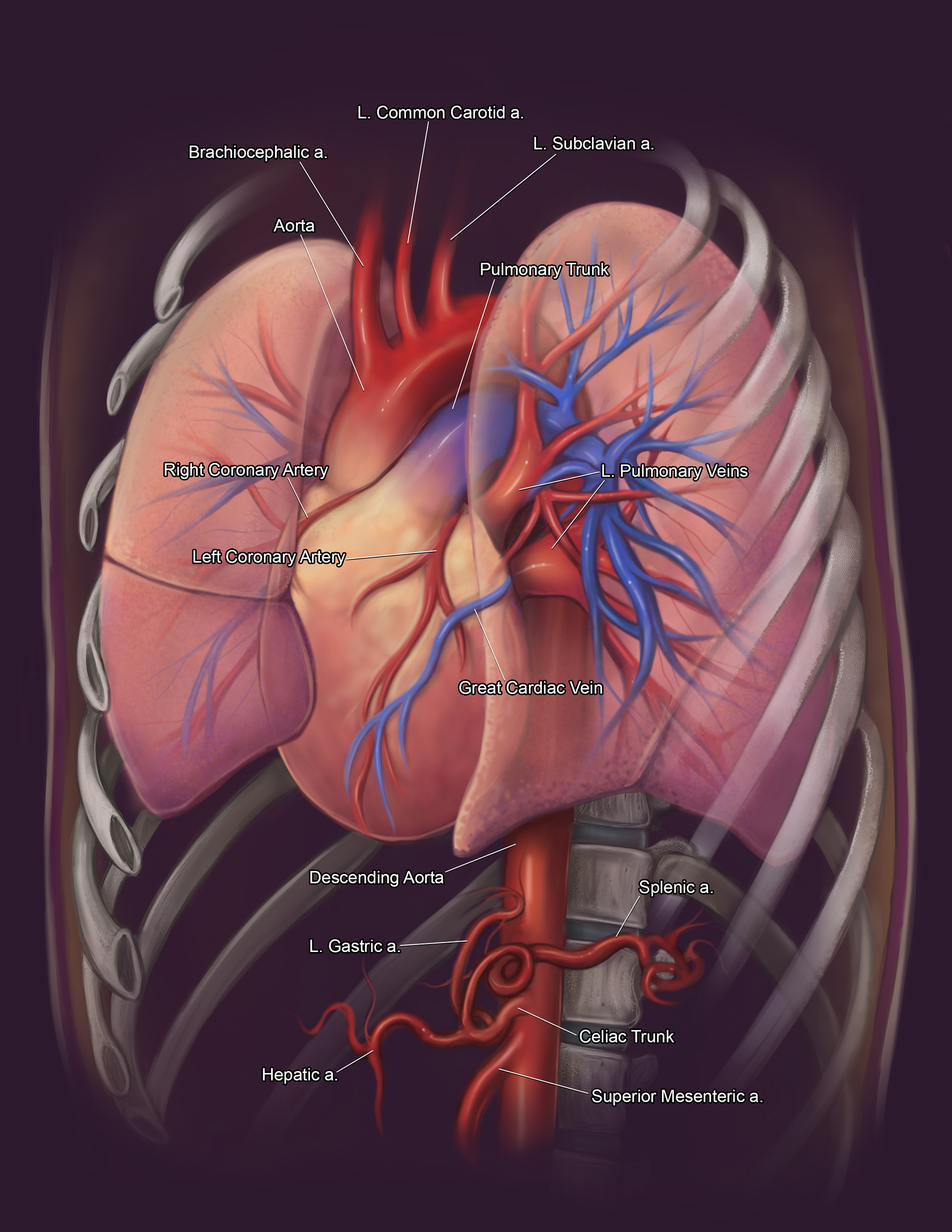
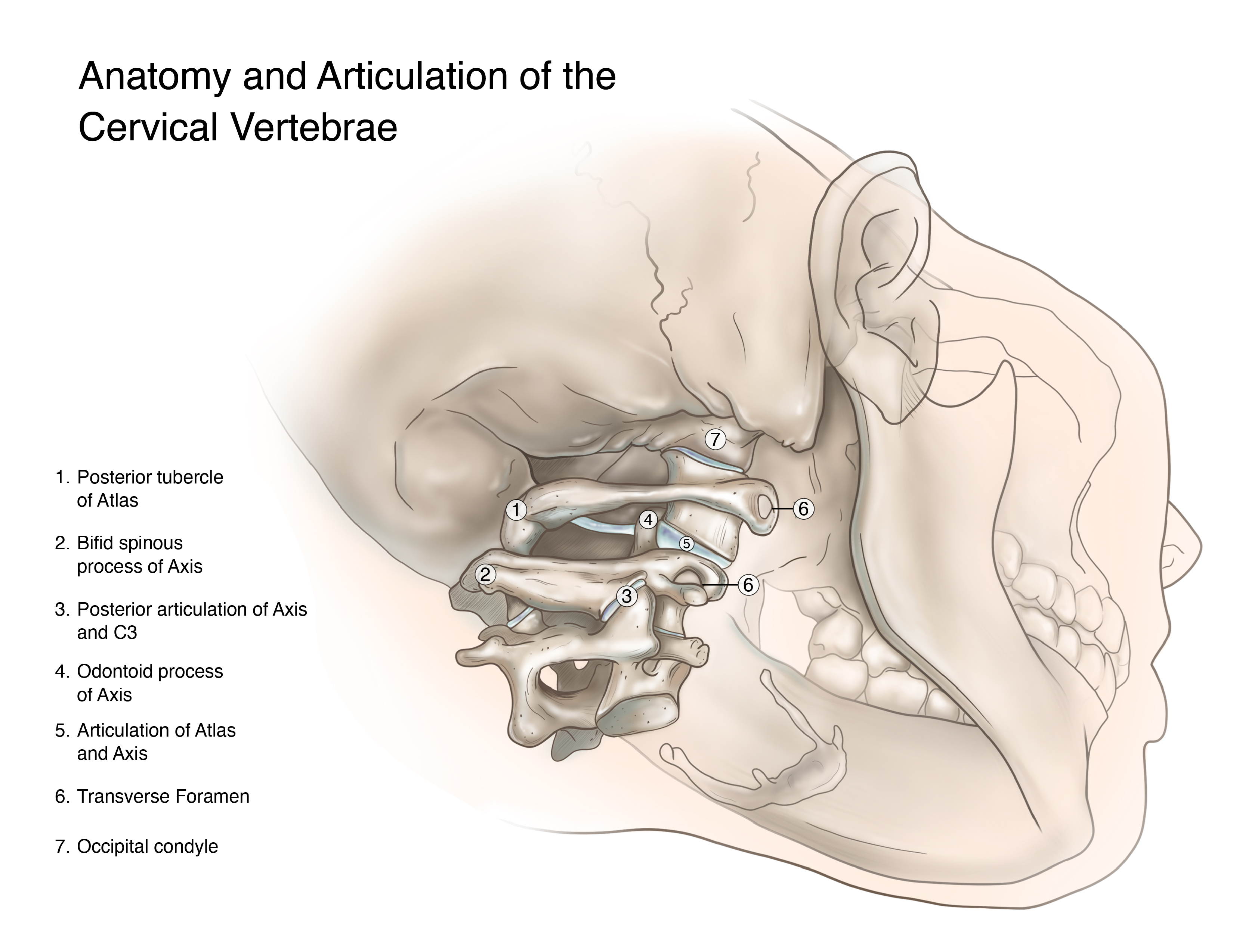
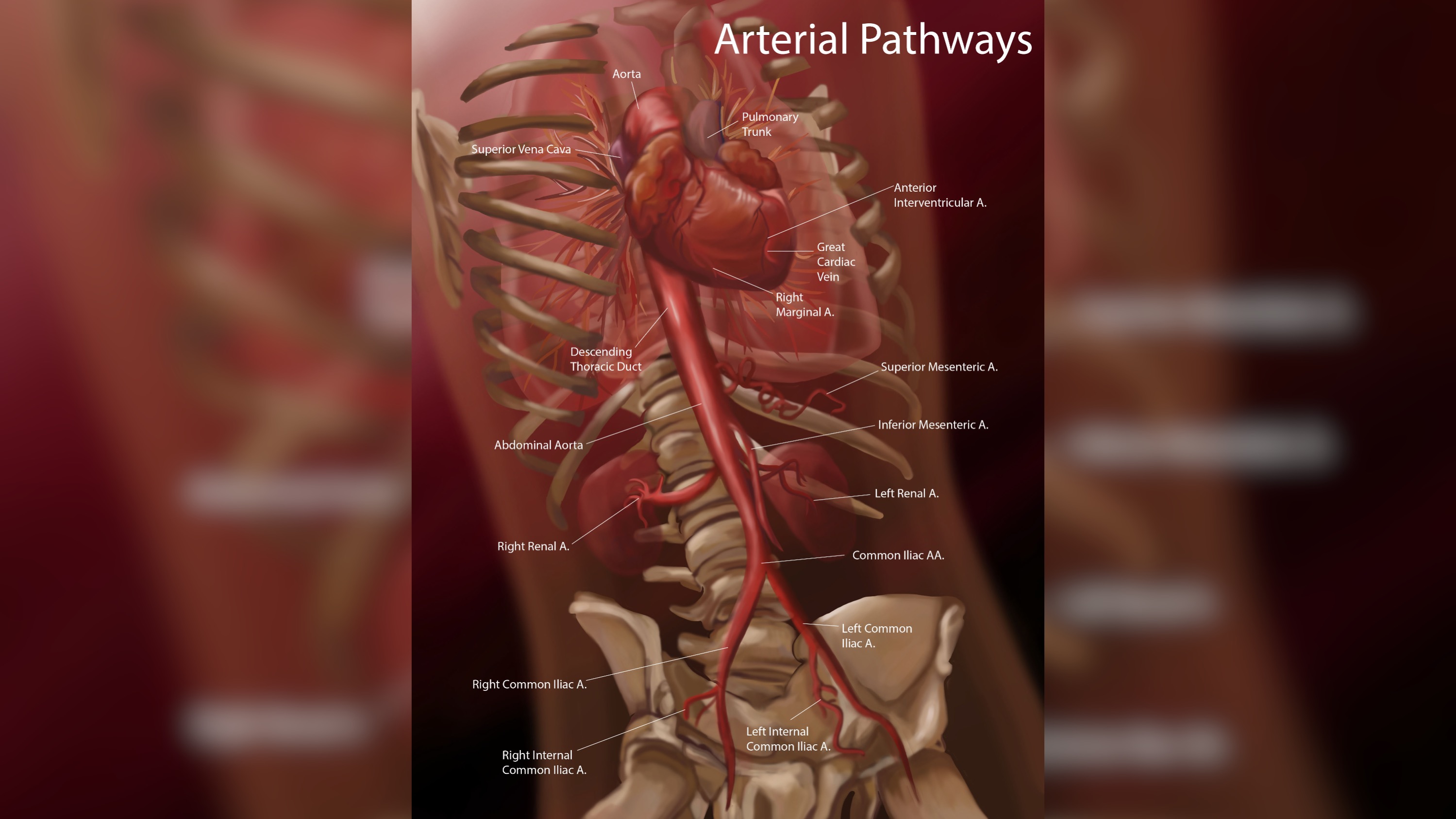
The Scientific Visualization class teaches students to locate and visualize data from various imaging modalities at the anatomic, cellular and molecular levels. They reconstruct the data in 3D and either convert the data directly into 3D models or export references for creating 2D images. In this case, students used CT and MRI data from actual patients, reconstructed the data in 3D using a program called Horos and exported reference images to draw the anatomy in 2D using Photoshop. The Photoshop images are highly accurate because they are based on references from actual, living patients.
Featured scientific illustration by Kiara Hoefsmit
By Masako Moyer
By Xavier Williams

